
Signs of this syndrome are severe pain with stretch of the big toe, loss of sensation in the foot or pain out of proportion to the injury itself. Eventually, the muscle will die if this goes untreated. This occurs when the pressure in the leg gets too high for blood and oxygen to circulate. Patients with tibia plateau fractures are at risk for a serious condition called compartment syndrome.
#OCCULT TIBIAL PLATEAU FRACTURE SKIN#
Often, the bone tries to poke out of the skin or “tent.” If this is not corrected, the skin can die or the bone can eventually cut the skin. The doctor will look for any open wounds over the injury as these usually require surgery. Diagnosis of an injury to some blood vessels requires urgent surgery. Important nerves and blood vessels run next to this bone and can be injured when it breaks.

Physical examination is critical in the evaluation of these injuries. If this is disrupted, arthritis can occur. It is also covered with a layer of cartilage that allows the knee to glide smoothly. They require this bone to be strong and straight to function well. The ligaments and tendons around the knee all connect to the plateau. The tibia plateau is an important part of the knee joint because it supports your body weight as you walk, run and jump.
#OCCULT TIBIAL PLATEAU FRACTURE CRACK#
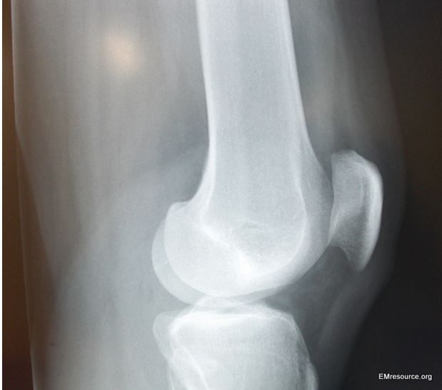
The tibia can be broken into many pieces or just crack slightly depending on the quality of bone and the type of injury. These fractures usually result from high energy injuries such as car accidents in younger patients and most often from falls in the elderly patient. All of these structures must be taken into account when diagnosing and treating these injuries. This is an injury that can involve the bone, meniscus, ligaments, muscles, tendons and skin around the knee. Tibial Plateau Fracture What is a Tibial Plateau Fracture?Ī tibial plateau fracture is a break of the larger lower leg bone below the knee that breaks into the knee joint itself. John Zebrack, MD General Orthopedic Surgery Jeffrey Webster, MD General Orthopedic Surgery Nichole Joslyn, MD Hand & Upper Extremity

Thomas Christensen, MD Hand & Upper Extremity James Christensen, MD Hand & Upper Extremity I think preoperative MRI is necessary to better treatment of fracture & treatment of periarticular soft tissue injury in tibial plateau fracture.Nikola Babovic, MD Hand & Upper Extremity CONCLUSION MRI can find additional fracture line or articular depression that can't be found in simple radiograph and gives more information about articular depression and soft tissue that is useful in surgical plans. There were 58 (85.3%) cases of soft tissue injury in MRI. There was no significant difference between MRI and simple radiograph of displaced bone fragment (p=0.168). It increased by 1.35 mm and it was meaningful statistically (p<0.05). Average depression of articular surface in simple radiograph was 2.93 mm and 4.28 mm in MRI. RESULTS There were 7 examples of Schatzker type change after MRI check. MATERIALS AND METHODS Compared MRI with simple radiograph about Schatzker classification, depression of articular surface and displacement of bone fragment from the 68 examples who checked MRI and we evaluated soft tissue injury around knee joint. Abstract PURPOSE To compare information about fracture type in MRI with simple radiograph in tibial plateau fractures and evaluate tibial plateau fractures type and accompanying soft tissue injury, and evaluate usefulness of MRI in tibial plateau fractures.


 0 kommentar(er)
